Opening
Rapid response call, 0200
- Ward 7, bed 12: “BP 78/40”
- You’re the first doctor there
Question
What are the first three things you’d do when you walk into the room?
What if the BP was 93/60?
- Does that change anything?
- What if the patient is 22? What if they’re 85?
Question
Is 93/60 “normal”? What else would you want to know?
By the end of this session you should be able to
- Recognise shock before the blood pressure falls
- Classify shock by mechanism using one equation
- Use a structured approach to the undifferentiated shocked patient: resuscitate while you work out the type
- Tell the types apart at the bedside, and know when to escalate
The Framework
What is shock?
- Inadequate tissue oxygen delivery:
- Almost always mediated by low blood pressure, but hypotension is the consequence, not the definition
One equation
- SV depends on preload, contractility and afterload
Question
If perfusion pressure is low, which terms could be responsible?
Where it can fail
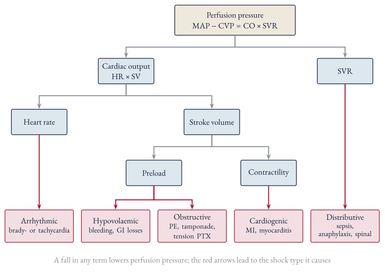
Four mechanisms
| Mechanism | Problem | Examples |
|---|---|---|
| Hypovolaemic | ↓ preload | Haemorrhage, GI losses, burns |
| Cardiogenic | ↓ contractility / rhythm | Acute MI, myocarditis, heart block |
| Obstructive | ↓ venous return / outflow | PE, tamponade, tension pneumothorax |
| Distributive | ↓ SVR | Sepsis, anaphylaxis, spinal injury |
A better mnemonic: PROVED?
- Pump
- Rhythm
- Obstructive
- Volume
- Endocrine
- Distributive
- ? Is it real?
Recognising Shock
Shock is a clinical diagnosis
- Haemodynamics: MAP < 65 or a big drop from baseline, shock index (HR/SBP) > 0.8
- Skin: cool knees and hands, mottling, CRT > 5 s
- Kidneys: urine output < 0.5 mL/kg/h
- Brain: agitation → delirium → drowsiness
- Blood: ↑ lactate, ↓ HCO₃⁻
Look at the knees
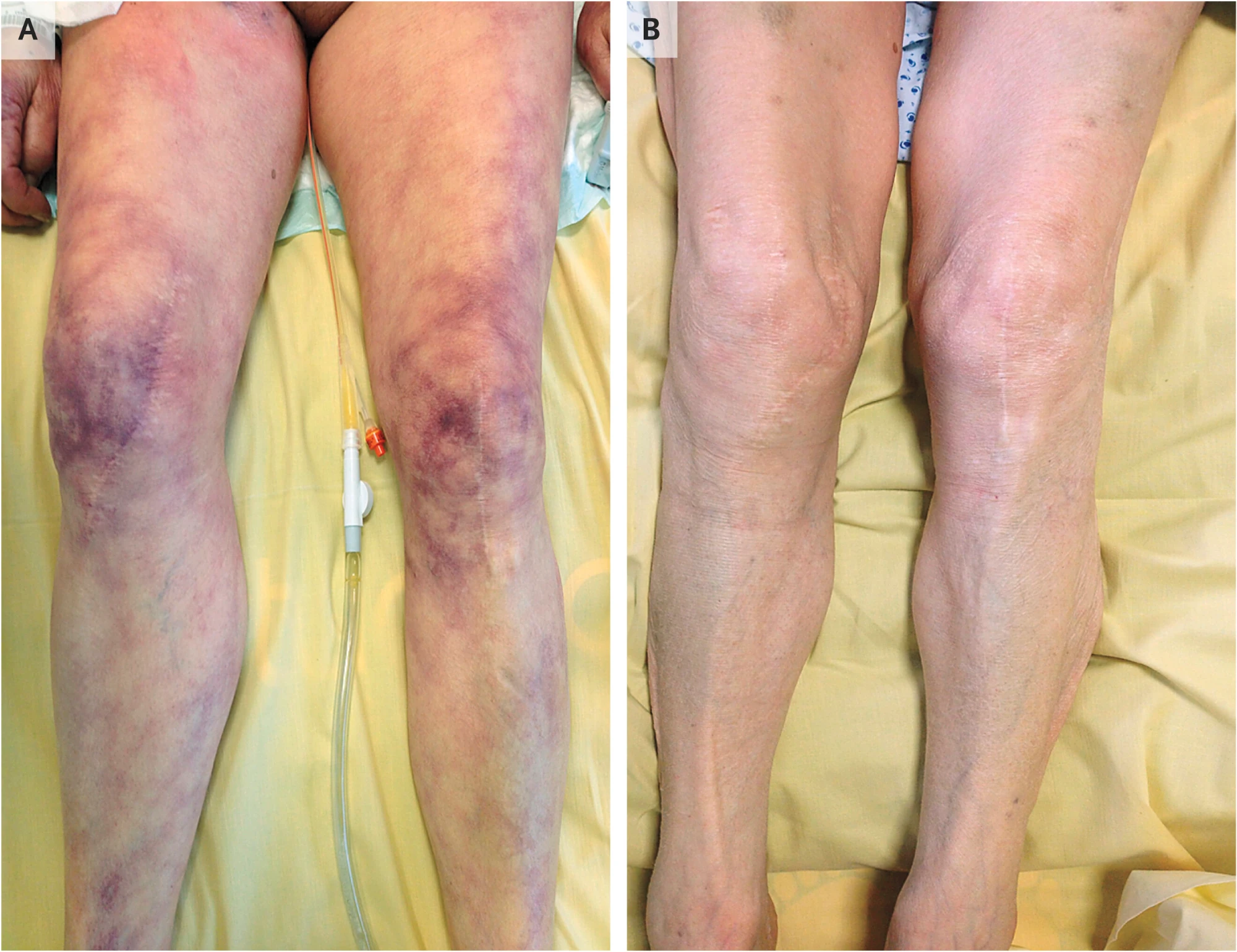
Contou D, de Prost N. Skin Mottling. N Engl J Med 2016;375:2187. doi:10.1056/NEJMicm1602055
Question
What’s the difference between A and B?
The Approach
Resuscitate while you differentiate
- Recognise it’s shock and call for help
- Primary survey: ABC, monitoring, exclude the immediate killers
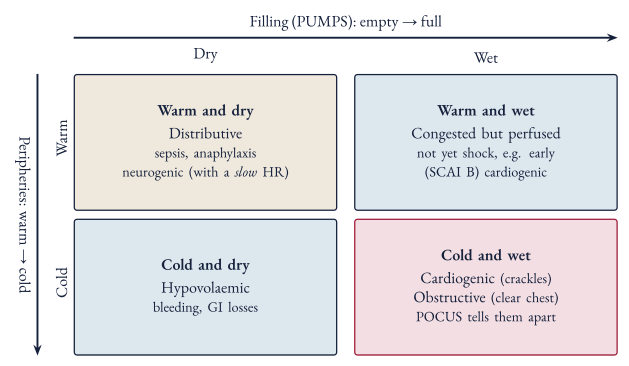
- Differentiate at the bedside: peripheries, heart rate, filling (PUMPS), POCUS
- Empirical resuscitation: fluid challenge, vasopressor, specific treatment
- Secondary survey: history, examination and investigations for the cause
- Reassess and escalate
The primary survey
- Call for senior help early
- A, B, C with monitoring on
- Exclude the things that kill quickly: tension pneumothorax, massive haemothorax, external haemorrhage, anaphylaxis, tamponade, massive PE
- IV access, bloods including cultures, ABG/VBG with lactate, 12-lead ECG
- Peri-arrest? Adrenaline 0.1 mg boluses to buy time while setting up an infusion
Three bedside questions
- Peripheries: warm or cold? (SVR)
- Heart rate: fast, slow or normal?
- Filling: full or empty?
Assessing filling: PUMPS
- Peripheral perfusion: capillary refill time, systolic BP, pulse pressure
- Urine output
- Mucous membranes and skin turgor
- Pressure in the neck: JVP
- Signs of volume overload: peripheral oedema, hepatojugular reflux, S3 gallop, lung crackles
Question
Which of these can you check in the first two minutes?
History that points to the type
| Ask about | Think |
|---|---|
| Chest pain, diaphoresis, known cardiac disease | Cardiogenic |
| Fevers, rigors, immunosuppression | Septic |
| Allergen exposure | Anaphylaxis |
| Trauma, blood loss | Hypovolaemic |
| Recent chest procedure, malignancy | Obstructive |
| Exogenous steroids, adrenal disease | Adrenal crisis |
Case 1: Mrs P
Mrs P, 78
Rapid response call, ward 7. Day 2 of an admission for a UTI, and the nurse says she’s “not herself”.
Ask for it
HR 118 · BP 82/45 · SpO2 94 · RR 26 · Temp 38.9
Warm, fast, empty: distributive
- Peripheries: warm, CRT 2 s → low SVR
- Heart rate: 118 → fast
- Filling (PUMPS): dry mucous membranes, JVP not visible, no signs of overload, urine output ≈ 0.17 mL/kg/h → empty
- With a fever and a known UTI, this is septic shock until proven otherwise
Sepsis and septic shock (Sepsis-3)
- Sepsis: life-threatening organ dysfunction due to a dysregulated host response to infection
- Organ dysfunction = increase in SOFA score ≥ 2
- Septic shock: sepsis plus both
- Hypotension requiring vasopressors
- Lactate > 2 mmol/L
Mrs P: what do you do first?
HR 118 · BP 82/45 · SpO2 94 · RR 26 · Temp 38.9
A bolus, then reassess
- MAP 67, CRT faster, a bit more alert
- That’s a fluid challenge: give it, then look at the patient again
- Is she still underfilled? Then another bolus, and reassess again
HR 106 · BP 96/52 · SpO2 95 · RR 23 · Temp 38.9
Two litres stat
- MAP up a little, but she’s now short of breath
- Crackles at both bases, SpO₂ 89%
- She’s 78: fluid overload is harmful too
- Give fluid as a challenge, and reassess after each one
HR 112 · BP 92/50 · SpO2 89 · RR 32 · Temp 38.9
Noradrenaline straight away
- Not wrong: start noradrenaline early, and peripherally is fine short-term
- But she’s dry as well as vasodilated, so she needs some fluid too
- Her urine output hasn’t picked up
HR 114 · BP 90/50 · SpO2 94 · RR 26 · Temp 38.9
Waiting for the bloods
- 40 minutes later: more confused, MAP 50
- You already had a lactate from the VBG
- Shock doesn’t wait for a lab result
- Antibiotics within 1 hour of recognising septic shock, after cultures
HR 132 · BP 74/38 · SpO2 91 · RR 32 · Temp 39.2
Fluids, done properly
- Give as a challenge: 250–500 mL over 10–15 min, then reassess
- 30 mL/kg within the first 3 hours for sepsis
- Balanced crystalloid (Hartmann’s, Plasma-Lyte) in most patients
Question
“Don’t we always give normal saline?”
Saline or balanced crystalloid?
- Balanced crystalloid is generally preferred in septic shock
- A conditional recommendation from the Surviving Sepsis Campaign, based on low quality evidence
- Normal saline is preferred in:
- Hypochloraemic alkalosis (e.g. vomiting, NGT losses)
- Hyperkalaemia
- Traumatic brain injury
- Meningitis
We’re bad at predicting who needs fluid
- Being fluid responsive doesn’t mean the patient needs fluid
- MAP and urine output lag behind and don’t reliably predict fluid responsiveness
- So give a bolus, then ask whether the patient actually got better:
- Capillary refill time and mottling
- Urine output
- Repeat lactate at 2 hours (aim for > 10% clearance)
- Mental state
An hour later: 2 L in, MAP 58
- Start noradrenaline; don’t wait for large fluid volumes
- Peripheral vasopressors are acceptable short-term through a large proximal vein
- Target MAP 65 (60–65 if aged ≥ 65)
- Escalate to ICU
HR 112 · BP 86/44 · SpO2 94 · RR 25 · Temp 38.7
Noradrenaline running
- MAP 71: at target
- Now reassess perfusion: CRT, mental state, urine output, repeat lactate
- ICU review for ongoing vasopressor needs
HR 102 · BP 104/54 · SpO2 95 · RR 22 · Temp 38.5
Case 2: Mr T
Mr T, 34
Motorbike vs car at 60 km/h. Ambulance handover in resus.
Ask for it
HR 124 · BP 108/84 · SpO2 97 · RR 28
Cold, fast, empty: hypovolaemic
- Peripheries: cool to the knees, CRT 4 s → vasoconstricted
- Heart rate: 124 → fast
- Filling (PUMPS): narrow pulse pressure, JVP not visible → empty
- In trauma, that’s haemorrhage until proven otherwise
The shock index
-
0.8 is elevated
- Mr T: 124 ÷ 108 ≈ 1.15
- Also look at his pulse pressure: 108 − 84 = 24 mmHg, which is narrow
Where’s the blood? SCALPeR
- Scalp / Street
- Chest
- Abdomen
- Long bones
- Pelvis
- Retroperitoneum
Question
How would you look for each at the bedside in the first 10 minutes?
Looking for it at the bedside
| Site | How |
|---|---|
| Scalp / street | Ambulance handover (blood at the scene), look at the scalp and under dressings |
| Chest | Examine, CXR, eFAST for haemothorax |
| Abdomen | Examine, eFAST for free fluid (Morison’s pouch first) |
| Long bones | Examine for deformity, X-ray |
| Pelvis | Pelvic X-ray, binder if suspected |
| Retroperitoneum | Hard to find at the bedside: POCUS isn’t sensitive, needs CT |
Mr T: what now?
HR 128 · BP 102/82 · SpO2 97 · RR 28
Two litres of crystalloid
- BP rose for ten minutes, then fell further
- Now 35.4 °C, and his INR is 1.6
- Crystalloid carries no oxygen and no clotting factors
- Bleeding patients need blood, with restricted volume replacement
HR 136 · BP 84/62 · SpO2 96 · RR 30 · Temp 35.4
Major haemorrhage protocol
- Blood products in a ratio no lower than 2:1:1, through a warmer
- TXA within 3 hours of injury
- Splinting the femur and getting him to definitive haemorrhage control
- SBP 92: no need to push higher until the bleeding is stopped
HR 112 · BP 92/64 · SpO2 98 · RR 24
A pressor for Mr T
- The BP looks better…
- …but he’s still bleeding and still empty: HR 130, pulse pressure 24
- Vasopressors aren’t the answer to an empty tank
- Noradrenaline only if restricted fluid can’t hold the target
HR 130 · BP 112/88 · SpO2 97 · RR 28
Off to CT
- Halfway through the scan, the radiographer calls: BP 76/58
- He’s in a corridor with no blood running
- Resuscitate and control the bleeding first; CT is for once he’s stable
HR 142 · BP 76/58 · SpO2 95 · RR 32
Managing haemorrhagic shock
- Stop the bleeding: that’s the definitive treatment
- Permissive hypotension: target SBP 80–90 until bleeding is controlled
- Not in TBI: keep MAP ≥ 80
- Activate the major haemorrhage protocol
- Tranexamic acid within 3 hours: 1 g over 10 min, then 1 g over 8 h
- Noradrenaline only if restricted fluid can’t hold the target
The major haemorrhage protocol
- Blood, not crystalloid
- RBC:FFP:PLT no lower than 2:1:1
- At least 1 FFP for every 2 RBC
- At least 1 adult unit of platelets for every 8 RBC
- Fibrinogen 3–4 g (cryoprecipitate or concentrate)
- Keep warm (≥ 35 °C), calcium up (iCa ≥ 1), and recheck bloods every 4 units
Case 3: Mr K
Mr K, 66
ED. Two hours of central chest pain, and he’s sweaty.
Ask for it
HR 108 · BP 84/60 · SpO2 89 · RR 30 · PVCs occasional
Cold, fast, full
- Peripheries: cold, mottled → vasoconstricted, low output
- Heart rate: 108 → fast
- Filling (PUMPS): JVP up, crackles, oedema → full
- With chest pain and ST elevation, cardiogenic shock is most likely…
- …but obstructive shock is cold and full too
Mr K: what next?
HR 110 · BP 82/58 · SpO2 88 · RR 30 · PVCs occasional
A bolus for Mr K
- No change in BP
- Crackles now to the mid zones, SpO₂ 82%
- He was already full: this is the fluid you can’t take back
HR 116 · BP 82/58 · SpO2 82 · RR 36
Noradrenaline, then dobutamine
- MAP up first with noradrenaline, then dobutamine for perfusion
- Right drugs, right order
- But nobody has looked at the heart yet. Is it really pump failure?
HR 104 · BP 98/64 · SpO2 91 · RR 28
POCUS first
What does the probe show?
- Pump failure: this is cardiogenic shock
What if it had looked like this?
| Cause | POCUS | Treatment |
|---|---|---|
| Massive PE | Dilated RV, McConnell’s sign | Thrombolysis (alteplase 100 mg: 10–20 mg bolus, rest over 2 h) |
| Tamponade | Effusion, RV diastolic collapse | Pericardiocentesis |
| Tension pneumothorax | No lung sliding | Needle decompression |
- Obstructive shock: cold and full, but the heart muscle isn’t the problem
- Fluids and pressors only buy time; the obstruction has to be relieved
RUSH, pump, tank, pipes
Overview of RUSH Exam
- Heart: LV function, RV dilation, pericardial effusion/tamponade
- IVC: size and collapsibility (volume responsiveness)
- Lungs: B-lines (pulmonary oedema), pneumothorax (absence of sliding)
- Abdomen: free fluid (haemoperitoneum, ruptured AAA)
- Aorta: AAA
- Lower limbs: DVT (if PE suspected)
Can use the mnemonic HI-MAP (Heart, IVC, Morrison’s pouch, Aorta, Pneumothorax) or alternatively go in the approach of pump (heart), tank (IVC, Morison’s pouch), and pipes (aorta, DVT)
Probably not
Only give fluid if all of these are true:
- End-organ perfusion is inadequate
- No pulmonary congestion (e.g. no B-lines)
- The overall picture suggests true hypovolaemia
- Dynamic indices suggest he’s fluid responsive
Cold and wet
Managing cardiogenic shock
- 12-lead ECG and POCUS early
- Support the lungs: CPAP/BiPAP
- Noradrenaline first to restore MAP, and keep an eye on the DBP (≥ 60 mmHg) for coronary perfusion
- Then dobutamine if still poorly perfused (cold, mottled)
- Adrenaline alone is reasonable as a short-term bridge
- Treat the cause: revascularisation for ACS
- Escalate early: ICU and cardiology
Putting It Together
Same three questions, different answers
| Peripheries | Heart rate | Filling | Clue | |
|---|---|---|---|---|
| Mrs P: distributive | Warm | Fast | Empty | Fever, source |
| Mr T: hypovolaemic | Cold | Fast | Empty | Trauma, blood loss |
| Mr K: cardiogenic | Cold | Fast | Full | Chest pain, crackles |
| Obstructive | Cold | Fast | Full | Often a clear chest; POCUS |
| Neurogenic | Warm | Slow | Empty | Spinal injury |
Don’t miss these
| Situation | Clue | First move |
|---|---|---|
| Anaphylaxis | Exposure, urticaria, wheeze | Adrenaline 0.5 mg IM |
| Adrenal crisis | Steroid use, ↓Na ↑K | Hydrocortisone 100 mg IV + saline |
| Neurogenic | Spinal injury, warm + bradycardic | Exclude haemorrhage; fluid, then vasopressor (MAP 85–90) |
| Bradycardic | Heart block on ECG | Atropine → isoprenaline, pacing |
| ”Is it real?” | Patient looks well | Recheck the cuff or line |
Wrap-Up
Your first 15 minutes
- Recognise it and call for help
- Primary survey: ABC, monitoring, access, VBG with lactate, ECG
- Differentiate: peripheries, heart rate, filling (PUMPS), POCUS
- Resuscitate: fluid challenge, pressor, inotrope, blood, or fix the obstruction
- Secondary survey: history, examination, investigations
- Reassess, and escalate
Take-home
- Shock is a failure of oxygen delivery; BP falls late
- tells you where to look
- Warm vs cold, fast vs slow, full vs empty (PUMPS)
- Resuscitate while you differentiate, and reassess after everything you give
- More than one type can co-exist (e.g. sepsis + hypovolaemia)
- Full notes: Shock
Sources: guidelines and trials
- Singer M, et al. The Third International Consensus Definitions for Sepsis and Septic Shock (Sepsis-3). JAMA. 2016;315(8):801–10
- Surviving Sepsis Campaign: International Guidelines for Management of Sepsis and Septic Shock 2026. Crit Care Med. 2026
- Asfar P, et al. High versus low blood-pressure target in patients with septic shock (SEPSISPAM). N Engl J Med. 2014;370:1583–93
- ARISE FLUIDS Investigators. Vasopressors or fluids in early septic shock. N Engl J Med. 2026. doi:10.1056/NEJMoa2516225
- National Blood Authority. Patient blood management guideline for adults with critical bleeding, v2.0. 2025
- Rossaint R, et al. The European guideline on management of major bleeding and coagulopathy following trauma: sixth edition. Crit Care. 2023;27:80. doi:10.1186/s13054-023-04327-7
- CRASH-2 trial collaborators. Effects of tranexamic acid in trauma patients with significant haemorrhage. Lancet. 2010;376:23–32. doi:10.1016/S0140-6736(10)60835-5
- NSW Agency for Clinical Innovation. Management of cardiogenic shock
Sources: reviews and further reading
- Thiele H, Hassager C. Cardiogenic shock. N Engl J Med. 2025;394(1):62–77. doi:10.1056/NEJMra2312086
- Puttur Rajkumar K, et al. Blood pressure goals in critically ill patients. Methodist DeBakey Cardiovasc J. 2023;19(4):24. doi:10.14797/mdcvj.1260
- Queensland Spinal Cord Injuries Service. Spinal and neurogenic shock
- Contou D, de Prost N. Skin mottling. N Engl J Med. 2016;375:2187. doi:10.1056/NEJMicm1602055 (mottling image)
- The POCUS Atlas: RUSH, undifferentiated hypotension
- Deranged Physiology: Approach to the undifferentiated shock patient
- LITFL: Shock · ICU One Pager: Undifferentiated shock
- Figures drawn for this tutorial; full notes and references in Shock